The Digital Fish Library
Research Collaborations
Learn more about that various research projects involving the implementation of high-field anatomical magnetic resonance imaging (MRI) to the study of marine organisms and comparative morphology of fishes.
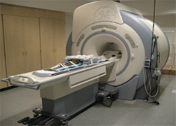 3T Scanner
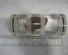 Fish Holder
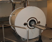 7T Scanner
-
1. Shark Olfaction +
Images
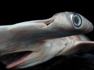 Hammerhead specimen
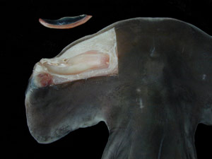 Disection showing the rosette organ
Project Description
Sharks possess among the largest olfactory organs of any vertebrate and hammerhead sharks in particular have a greatly expanded olfactory rosette. Their large olfactory organs and acute sensitivity have contributed to their reputation as swimming noses. However, very few studies have actually tested the threshold sensitivity of sharks to odor stimuli. We will employ functional MRI to image the olfactory organs of sharks as they are exposed to a variety of odorants to determine how low a concentration the sharks are actually able to detect. The functional data will be combined with high resolution anatomical data to better understand the form and function of the olfactory organ in the sharks.
Collaborators
Stephen M. Kajiura
Elasmobranch Research Laboratory at Florida Atlantic University
Tricia Meredith
Elasmobranch Research Laboratory at Florida Atlantic University
Lawrence Frank
University of California, San Diego -
2. Shark Brain Morphology +
Images
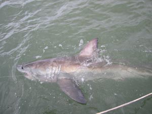 This juvenile great white was caught, tagged, and released as part of an ongoing tracking study
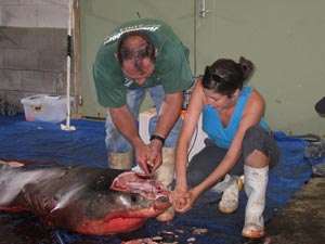 This great white shark was found drowned in a gill net and donated for scientific study
Project Description
Chondrichthyans (cartilaginous fishes) occupy a basal position in vertebrate evolution and offer a relatively unexplored opportunity to study the evolution of vertebrate brains. Our study examines the brain morphology of elasmobranchs (sharks, skates, and rays) in relation to both phylogeny and ecology. There is significant variation in both brain size and complexity across cartilaginous fish species. Using MRI, we will assess relative brain size and the relative size of five major brain areas (telencephalon, diencephalon, mesencephalon, cerebellum, and medulla) in a range of chondrichthyan species. This project aims to work with other members of the DFL project to contribute to the master specimen digital database.
Collaborators
Kara Yopak
University of California, San Diego
Lawrence Frank
University of California, San Diego
Glenn Northcutt
Scripps Institution of Oceanography -
3. Ponyfish (Leiognathidae) Light Organs +
Images
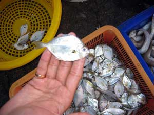 Freshly caught ponyfish
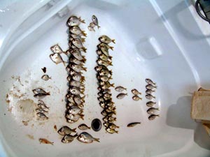 Specimens sorted out in a bathtub
 Project description
We are interested in studying Leiognathidae (ponyfish) because they provide an extraordinary and unmatched biological system that combines dramatic sexual dimorphism and photic signaling. The light organ system in these fishes differs greatly between species and sexes, but is seldom studied and not very well understood. As our group works out phylogenetic relationships between these fishes, we will be able to better understand how this light organ system evolved. Part of understanding the relationships requires collecting new material from the different parts of the world these species are found. While collecting, we found that many species were undescribed and most of the taxonomy was incorrect. Unfortunately, much of the original material used to describe these species (type specimens) is lost or in poor condition. With MRI we are able to find important characters in this rare material that would otherwise be concealed to us. We can see more structural detail than would be possible through dissection, and we can view the entire system in situ. This significantly improves our ability to identify ponyfish clades and helps us better understand the evolution of the light organ system.
Collaborators
John Sparks
American Museum of Natural History
Prosanta Chakrabarty
American Museum of Natural History
Lawrence Frank
University of California, San Diego -
4. Cranial Endothermy in Opah (Lampris guttatus) +
Images
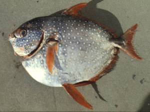 Opah specimenn
Project description
Cranial endothermy is the ability of certain fishes to maintain the temperature of the cranial region (eye and brain) at a temperature significantly higher than the surrounding water temperature. Cranial endothermy has evolved by convergence in billfishes, the butterfly mackerel, tunas, and lamnid sharks. The purpose of this project is to document cranial endothermy in another teleost fish species, the opah or moonfish (Lampris guttatus). Like all other cranial endotherms, this species experiences rapid changes in seawater temperature as it moves vertically within the water column. MRI allows us to visualize the three-dimensional arrangement of the heat source and the heat retention mechanisms required for cranial endothermy in L. guttatus. This study will contribute to understanding the evolution of endothermy in fishes and the physiological ecology of this unique fish.
Methods
T1-weighted 3D spoiled gradient recalled echo acquisition with image and segmentation analyses
Collaborators
Kathryn Dickson
California State University Fullerton
Rosa Runcie
California State University Fullerton
Donald Hawn
National Marine Fisheries Service, Pacific Islands Fisheries Science Center, Honolulu, Hawaii
Heidi Dewar
National Marine Fisheries Service, Southwest Fisheries Science Center, La Jolla, CA
Lawrence Frank
University of California San Diego -
5. Red Muscle in shortfin mako shark (Isurus oxyrinchus) and salmon shark (Lamna ditropis) +
Project description
To differentiate muscle fiber types and quantify the amounts of slow, red aerobic muscle in the shortfin mako shark (Isurus oxyrinchus) and the salmon shark (Lamna ditropis)
Methods
T1-weighted 3D spoiled gradient recalled echo acquisition with image and segmentation analyses.
Investigators
Cameron Perry, Daniel Cartamil, Diego Bernal, Chugey A. Sepulveda, Rebecca J. Theilmann, Jeffrey Graham, Lawrence R. Frank - University of California, San Diego
-
6. Coral Complexity +
Images
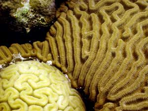 Brain coral
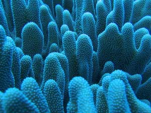 Soft coral
Videos
Animations of rendered coral MR data

Project description
We are interested in how topographic complexity contributes to the biodiversity and habitat complexity of benthic communities, especially coral reefs. Coral reefs represent one of the roughest structures in the marine environment. This roughness is a significant component of the high degree of habitat complexity associated with reefs. Various studies have investigated reef rugosity at discrete spatial scales, ranging from kilometers down to millimeters. However, to date, few studies have addressed fine-scale roughness (<1 cm). MR images allow us to construct high-resolution 3D surface models of individual corals, which we use to estimate their roughness on spatial scales from 200 um to 1 cm in terms of fractal dimension.
Collaborators
David Zawada
US Geological Survey
Forest Rohwer
San Diego State University
Lawrence Frank
University of California, San Diego